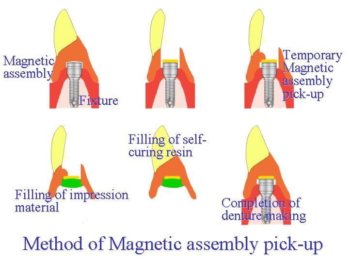
Ten years have passed since the clinical introduction of magnetic attachments. The clinical efficacy of magnetic attachments has been assessed during this period and new magnetic attachments are currently being evaluated. One application is the use of magnetic attachments for dental implants. Implant dentures with magnetic attachments differ from dentures retained by natural dental roots that have a periodontal membrane to allow physiologic movement. A keeper, which is a component of the magnetic assembly, is attached to an implant body that has been rigidly placed in alveolar bone. The abutment tooth must therefore be treated differently in terms of key supports and retentions that are integral in the design of partial dentures. In this study, a magnetic attachment developed for implants (MagFit-IP, Aichi Steel Corp., Japan) was used as a retention device in the maxillary right and left cuspid position in a subject with an edentulous maxilla, and a horse-shoe plate type denture was subsequently fitted. The clinical techniques involved in preparation, together with relevant findings, are reported here.
Case Summery Subject: 50-year-old male Chief
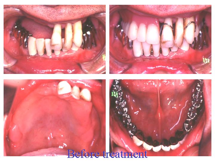
complaint: Masticatory disturbance Medical History: The subject had a maxillary partial denture fitted in order to restore lost teeth from the maxillary right central incisor to the second molar and from the maxillary left first premolar to the second molar. A full bridge was placed in the mandible. Although the prosthetic restoration functioned favorably afterwards, the subject recently complained of mobility of the remaining maxillary central incisor, lateral incisor, and cuspid as well as denture incompatibility due to resorption of the alveolar ridge. In addition, the subject expressed strong dissatisfaction with denture fit in the palatal region.
Treatment planning
There was significant alveolar ridge
resorption around the maxillary right central incisor, lateral
incisor, and cuspid. This was determined
to be an M3 category and could not be saved. The teeth were
extracted and two implants (Sterioss, Yoshida Dental, Japan)
were placed in the right and left cuspid positions. Six months
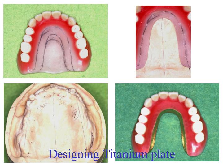
later, secondary surgery was performed and a horse-shoe plate
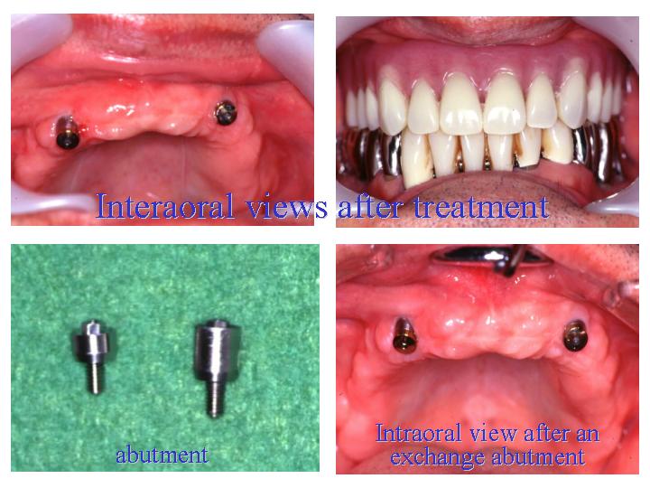
type titanium denture was prepared. However, an abutment of appropriate length
could not be found for the right-side implant, causing it to project slightly. The
gingival margin was lowered by brushing the gingiva and the
abutment was replaced with a smaller size after six months.
By
replacing the 5.5-mm
abutment with a 3-mm
abutment, a more favorable gingiva and fixture configuration was
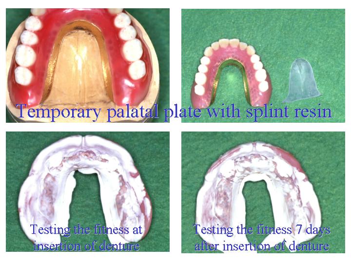
achieved. The labial flange of the denture could be lowered due
to use of the magnetic attachment,
which
resulted
in improved fit. The following benefits were obtained by placing
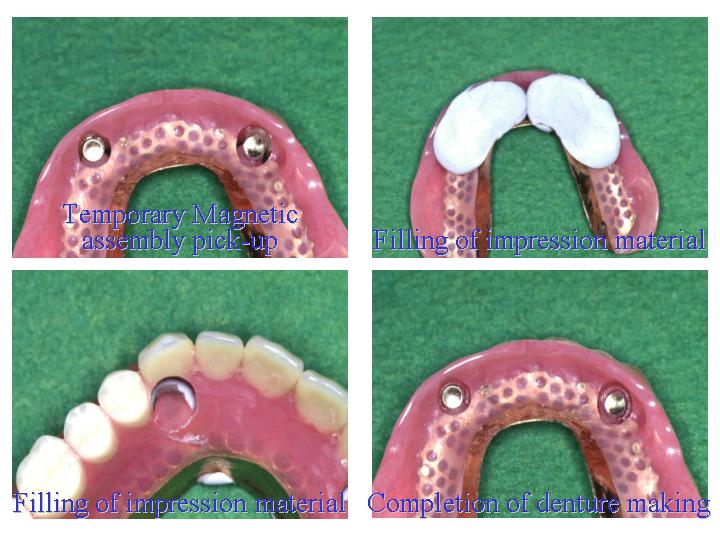
implants and using a magnetic attachment; 1.
By using a plate-type denture, retention was
achieved
with only two implants. 2.
The
palate remained
free. 3.
Reduction
of the labial flange was achieved. 4.
Effective
relief around the implants was achieved.
Both the
denture and implant have performed favorably for
16 months and the patient has expressed satisfaction with the
treatment.
|